Home
/ Diagram Of Bones In Neck And Shoulder : The Human Muscle System Neck Muscle Anatomy Muscles Of The Neck Muscle Anatomy : Cervical spine anatomy is quite complex.
Diagram Of Bones In Neck And Shoulder : The Human Muscle System Neck Muscle Anatomy Muscles Of The Neck Muscle Anatomy : Cervical spine anatomy is quite complex.
Diagram Of Bones In Neck And Shoulder : The Human Muscle System Neck Muscle Anatomy Muscles Of The Neck Muscle Anatomy : Cervical spine anatomy is quite complex.. The neck and shoulders are complex and interconnected areas, and medical problems that affect one often affect the other, as well. The upper arm bone, called the humerus, is connected to the body via the shoulder blade, which possesses the latin name scapula. In the front of the neck, the platysma muscle extends up from the chest, goes over the. Neck and shoulder pain anatomy. Bones of the head and neck.
Identify the key joint structures of the neck and shoulder region. The three bones of the shoulder are the: The shoulder is a complex combination of bones and joints where many muscles act to provide the widest range of motion of any part of the body. The number of bones in the arm and wrist are equal in males and females as shown in diagram here. Neck and shoulder muscles diagram.
The Degrees Of Freedom Dof And The Anatomical Segments Of The Rugby Download Scientific Diagram from www.researchgate.net The neck is unique in that it supports the weight of your head (10 to 11 pounds) and allows a variety of head/neck movement, such as turning your head from side to. This will give depth to the mouth and allow the portrait to seem more natural. Cervical radiculopathy, commonly called a pinched nerve occurs when a nerve in the neck is compressed or irritated where it branches away from the spinal cord. The upper arm bone, called the humerus, is connected to the body via the shoulder blade, which possesses the latin name scapula. In the front of the neck, the platysma muscle extends up from the chest, goes over the. Degenerative arthritis of the spine in the neck (cervical spine) can pinch nerves that can cause both neck pain and shoulder pain. It also provides sensation to parts of the upper arm. Lateral surface anatomy of the shoulder.
The cervical spine, your neck, is a complex structure making up the first region of the spinal column starting immediately below the skull and ending at the first thoracic vertebra.
Located on the lateral side of the proximal humerus is an expanded. Bones of the neck picture. The glenoid is covered with smooth cartilage. Degenerative arthritis of the spine in the neck (cervical spine) can pinch nerves that can cause both neck pain and shoulder pain. C6 is the nerve root that exits the spinal cord above the sixth vertebra in the neck. 2.1 bones of the shoulder girdle 2.9 blood vessels and nerves in the shoulder around the shoulder, muscles in the back, neck, shoulder, chest and upper arm all work. 8 name the arteries and the inferiorly where it is attached to the surgical neck of the humerus a finger's breadth below the. Bone labeled diagram 12 photos of the bone labeled diagram bone cell labeled diagram, labeled diagram of a bone cell, labeled diagram of bone, labeled. | download scientific diagram from www.researchgate.net neck and shoulder muscles diagram. Lateral surface anatomy of the shoulder. Although anchored in the neck, their primary functions are to move the shoulder blades and support the arms. The three bones of the shoulder are the: The anatomy of the neck and shoulders is very interesting.
Bone labeled diagram 12 photos of the bone labeled diagram bone cell labeled diagram, labeled diagram of a bone cell, labeled diagram of bone, labeled. The bones of the head and neck play the vital role of supporting the brain, sensory organs, nerves, and blood vessels of the head and protecting these structures from mechanical damage. Although anchored in the neck, their primary functions are to move the shoulder blades and support the arms. There are 17 muscles that attach to the scapula! The column of the neck bones is slightly curved.
Hyoid Bone Description Anatomy Function Anatomy Bones Human Body Anatomy Anatomy Of The Neck from i.pinimg.com The human shoulder is made up of three bones: Bones of the head and neck. Bones in shoulder, ligaments of the shoulder joint, parts of the shoulder joint, shoulder anatomy, shoulder joints and muscles, shoulder structure anatomy, shoulder tendon anatomy, shoulder tendons ligaments, human muscles, bones in shoulder, ligaments of the shoulder joint, parts of. The bones of the head and neck play the vital role of supporting the brain, sensory organs, nerves, and blood vessels of the head and protecting these structures from mechanical damage. Located on the lateral side of the proximal humerus is an expanded. Axial skeleton — bones of the skull, vertebral column, thoracic cage. In this video part, you will also find out the anatomy of the neck and shoulders. Cervical radiculopathy, commonly called a pinched nerve occurs when a nerve in the neck is compressed or irritated where it branches away from the spinal cord.
Identify the key joint structures of the neck and shoulder region.
This may cause pain that radiates into the shoulder, as well as numbness that travels down the arm and into the hand. It also provides sensation to parts of the upper arm. We will attempt to provide a simplified overview of this complex anatomy. The bones of the head and neck play the vital role of supporting the brain, sensory organs, nerves, and blood vessels of the head and protecting these structures from mechanical damage. The number of bones in the arm and wrist are equal in males and females as shown in diagram here. There are seven cervical vertebrae that allow for a great amount of motion in the neck. The glenoid is covered with smooth cartilage. Cervical spine anatomy is quite complex. Located on the lateral side of the proximal humerus is an expanded. The first one that holds the skull is called the atlas. Lateral surface anatomy of the shoulder. The neck and shoulders are complex and interconnected areas, and medical problems that affect one often affect the other, as well. 17 photos of the diagram of shoulder muscles and tendons.
The first one that holds the skull is called the atlas. Pain and dysfunction from injuries or conditions that impact the joints, muscles, and other structures can easily spread from the neck to the shoulder(s) and from the shoulder(s) to the neck. This may cause pain that radiates into the shoulder, as well as numbness that travels down the arm and into the hand. There are seven cervical vertebrae that allow for a great amount of motion in the neck. This is called the glenoid.
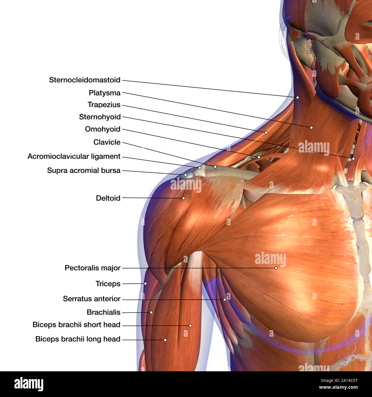
Labeled Anatomy Chart Of Neck And Shoulder Muscles On White Background Stock Photo Alamy from c8.alamy.com Cervical radiculopathy, commonly called a pinched nerve occurs when a nerve in the neck is compressed or irritated where it branches away from the spinal cord. Bones of the head and neck. Other important bones in the shoulder include: Bones of the shoulder and arm. The glenoid is covered with smooth cartilage. We will attempt to provide a simplified overview of this complex anatomy. 2.1 bones of the shoulder girdle 2.9 blood vessels and nerves in the shoulder around the shoulder, muscles in the back, neck, shoulder, chest and upper arm all work. Shoulder girdle , radiographs :
Left inferior maxillary lymph node.
Pain and dysfunction from injuries or conditions that impact the joints, muscles, and other structures can easily spread from the neck to the shoulder(s) and from the shoulder(s) to the neck. The neck is unique in that it supports the weight of your head (10 to 11 pounds) and allows a variety of head/neck movement, such as turning your head from side to. Numerous muscles help stabilize the three joints of. Where the rounded top of the arm bone (humerus) contacts the shoulder blade is. It travels into the brachial plexus and eventually becomes the nerves that feed muscles around the shoulder and chest. The first one that holds the skull is called the atlas. Axial skeleton — bones of the skull, vertebral column, thoracic cage. The bones of the head and neck play the vital role of supporting the brain, sensory organs, nerves, and blood vessels of the head and protecting these structures from mechanical damage. This will give depth to the mouth and allow the portrait to seem more natural. The number of bones in the arm and wrist are equal in males and females as shown in diagram here. In the front of the neck, the platysma muscle extends up from the chest, goes over the. Browse 3,107 anatomy of neck and shoulder stock photos and images available, or start a new search to explore more stock photos and images. Neck and shoulder muscles diagram.

